TM4000PlusIII
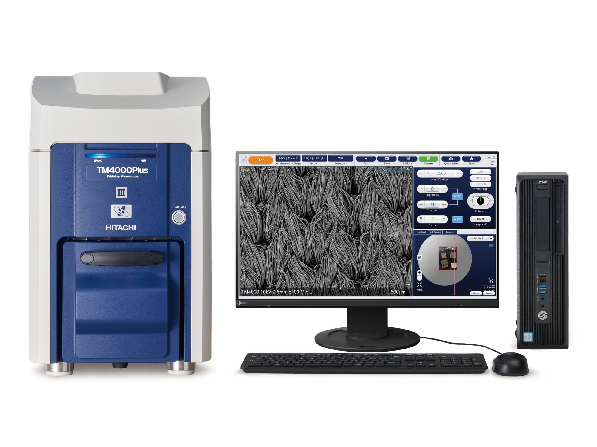
The new Tabletop Scanning Electron Microscope
High-performance tabletop SEM for flexible imaging needs in academia and industry.
- Automation for consistent and reliable imaging
- Optional elemental analysis
- Ideal for both conductive and non-conductive samples
- Well-resolved, high depth-of-focus and multiple-mode imaging
- Navigate with ease using optical camera integration
- Maintenance planning with filament monitoring
Overview
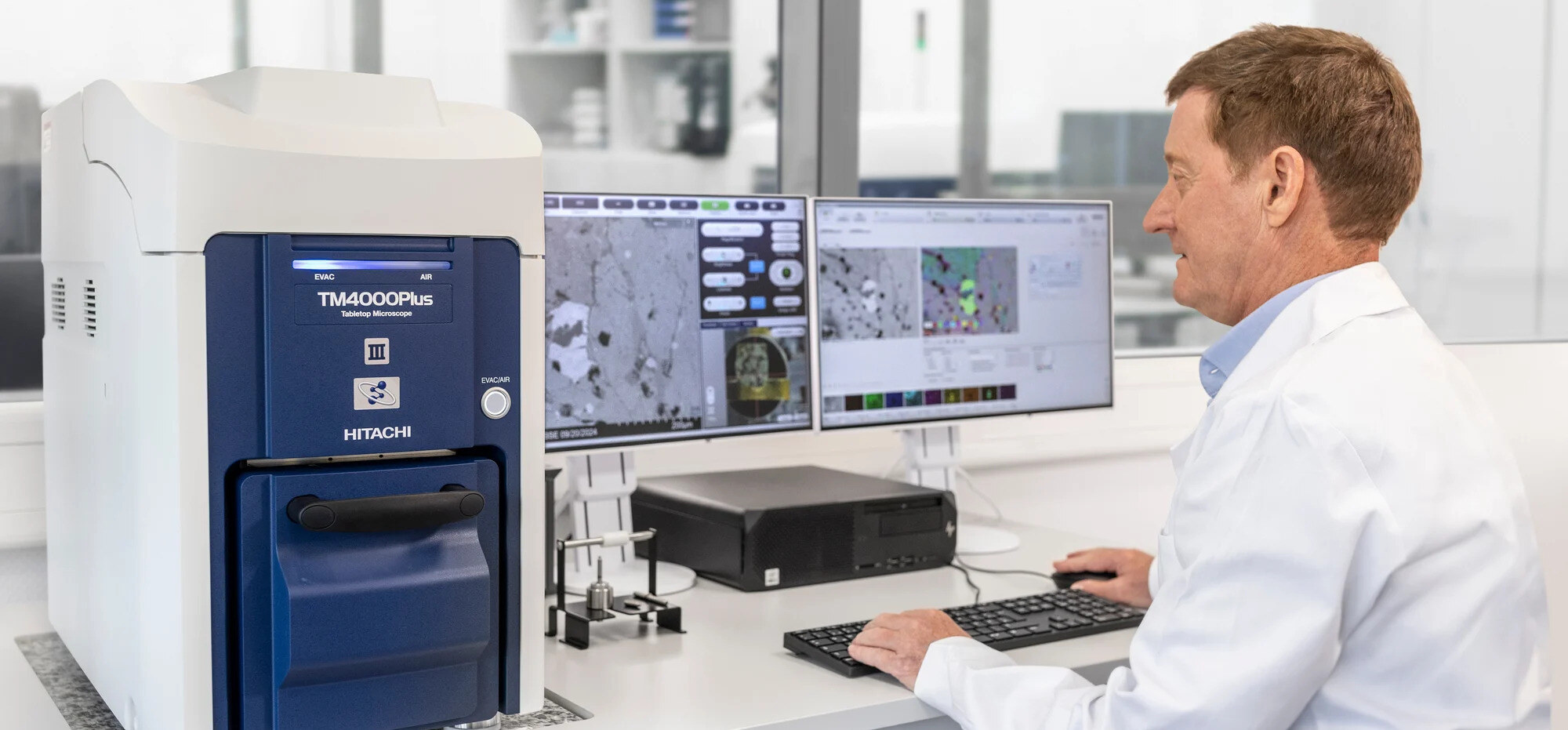
The Hitachi TM4000PlusIII scanning electron microscope offers a compact, user-friendly solution for imaging and elemental analysis. Designed for a range of users - from research labs to industrial quality control - this new tabletop SEM gives you reliable imaging capabilities without the costs and space requirements of a traditional SEM.
Why choose the TM4000PlusIII?
Whether you're a research scientist looking to deepen your understanding of materials or a quality control manager striving for consistent results, the TM4000PlusIII is built to meet your needs.
This microscope brings the power of scanning electron microscopy into an accessible, tabletop design. It combines ease of use with powerful features for imaging, navigation, and analysis.
With automated functions to streamline repetitive tasks, high signal-to-noise ratio for clear imaging, and an intuitive user interface, this SEM makes electron microscopy more approachable and productive then ever.

Features and benefits
-
Automation for consistent and reliable imaging
- Auto-focus, brightness, and contrast ensure consistent results across multi-user environments.
- Automated stage movement, magnification adjustments, and image capture simplify operation for inexperienced users.
- Optional tools like Multi-Zigzag, Python scripting, and EM-Flow Creator enable reproducible workflows without coding skills.

- Auto-focus, brightness, and contrast ensure consistent results across multi-user environments.
-
Well-resolved, high depth-of-focus and multiple-mode imaging
- Precise, contrast-rich imaging at 5, 10, or 15 kV.
- Backscattered and secondary electron detectors provide compositional, topographical, and surface details.
- Mixed output from both detectors delivers comprehensive imaging insights in a single image.

- Precise, contrast-rich imaging at 5, 10, or 15 kV.
-
Enhanced imaging flexibility and elemental analysis
- Optional integrated EDX for material composition analysis with 30 mm² or 60 mm² sensors.
- Switch seamlessly between imaging and elemental mapping for comprehensive sample studies.
- Compatible with AZtecLiveLite for efficient, high-throughput analysis.

- Optional integrated EDX for material composition analysis with 30 mm² or 60 mm² sensors.
-
Navigate with ease using optical camera integration
- Integrated high-resolution optical camera provides a zoomable map overview of samples.
- Pair optical images with SEM snapshots for precise navigation and seamless operation.

- Integrated high-resolution optical camera provides a zoomable map overview of samples.
-
Ideal for educational and non-conductive samples
- Low-vacuum functionality ensures clear imaging of delicate or non-conductive samples without extensive preparation.
- Suitable for educational settings, allowing students and early-career researchers to explore electron microscopy easily.

- Low-vacuum functionality ensures clear imaging of delicate or non-conductive samples without extensive preparation.
-
Maintenance planning with filament monitoring
- Tracks filament usage and projects remaining lifespan for proactive maintenance planning.
- Ensures uninterrupted workflows and minimizes downtime in labs with multiple users.

- Tracks filament usage and projects remaining lifespan for proactive maintenance planning.
Applications gallery

White metal
Acceleration voltage: 20kV, Magnification: 2,000x

Corroded copper wire
Acceleration voltage: 10kV, SE signal

STEM observation of grease thickener
Magnification: 9,000x

STEM observation of grease thickener
Magnification: 10,000x

Rat blood vessels (deparaffinized sections)
Acceleration voltage: 15kV, Magnification: 800x

Rat blood vessels (deparaffinized sections)
Acceleration voltage: 15kV, Magnification: 3,000x
Specifications
| Magnifications | 10x - 100,000x (Photographic), 25x - 250,000x (Monitor display) |
| Accelerating Voltage | 5 kV, 10 kV, 15 kV, 20 kV |
| Probe Current Mode | 5 steps |
| Image Signal | Backscattered, Secondary, Mixed |
| Vacuum Mode | Conductor: BSE, CL, Standard: BSE/SE/Mixed, Charge-up reduction: BSE/SE/Mixed |
| Stage Travel Range | X: 40 mm, Y: 35 mm |
| Camera Navigation System | Included |
| Filament Type | Pre-centered cartridge tungsten hairpin |
| Signal Detection System | High-Sensitivity 4-segment BSE, Low-Vacuum SE / CL |
| Auto Image-Adjustment | Auto start, focus, brightness |
| Image Data Saving | 2,560 x 1,920 pixels, 1,280 x 960 pixels, 640 x 480 pixels |
| Evacuation System | Turbo molecular pump, diaphragm pump |
| Operation Help Functions | Raster rotation, magnification presets, image shift |
| Safety Functions | Over-current protection, built-in ELCB |
Contact us
If you would like help choosing the best SEM for your needs, or if you’re looking for advice on how to optimise your microscopy, our experts are ready to assist. Contact us today to learn how the Hitachi TM4000PlusIII can enhance your lab’s capabilities.