
- Automation for consistent and reliable imaging
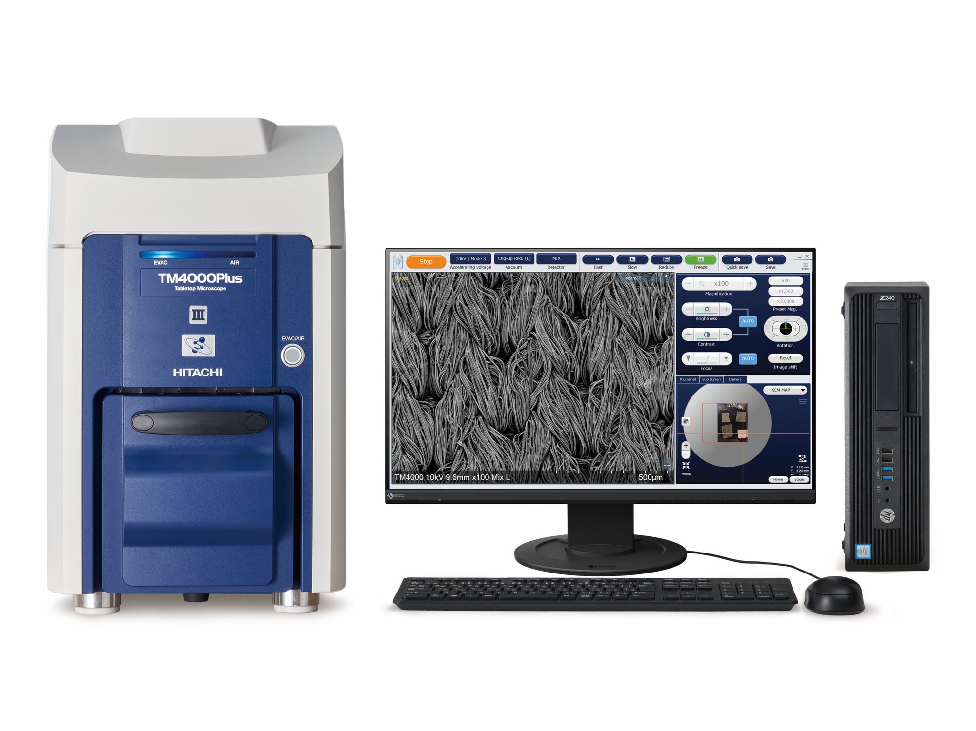
- Well-resolved, high depth-of-focus and multiple-mode imaging
- Enhanced imaging flexibility and elemental analysis
- Navigate with ease using optical camera integration
- Ideal for educational and non-conductive samples
- Maintenance planning with filament monitoring


White metal
Acceleration voltage: 20kV, Magnification: 2,000x

Corroded copper wire
Acceleration voltage: 10kV, SE signal

STEM observation of grease thickener
Magnification: 9,000x

STEM observation of grease thickener
Magnification: 10,000x

Rat blood vessels (deparaffinized sections)
Acceleration voltage: 15kV, Magnification: 800x

Rat blood vessels (deparaffinized sections)
Acceleration voltage: 15kV, Magnification: 3,000x