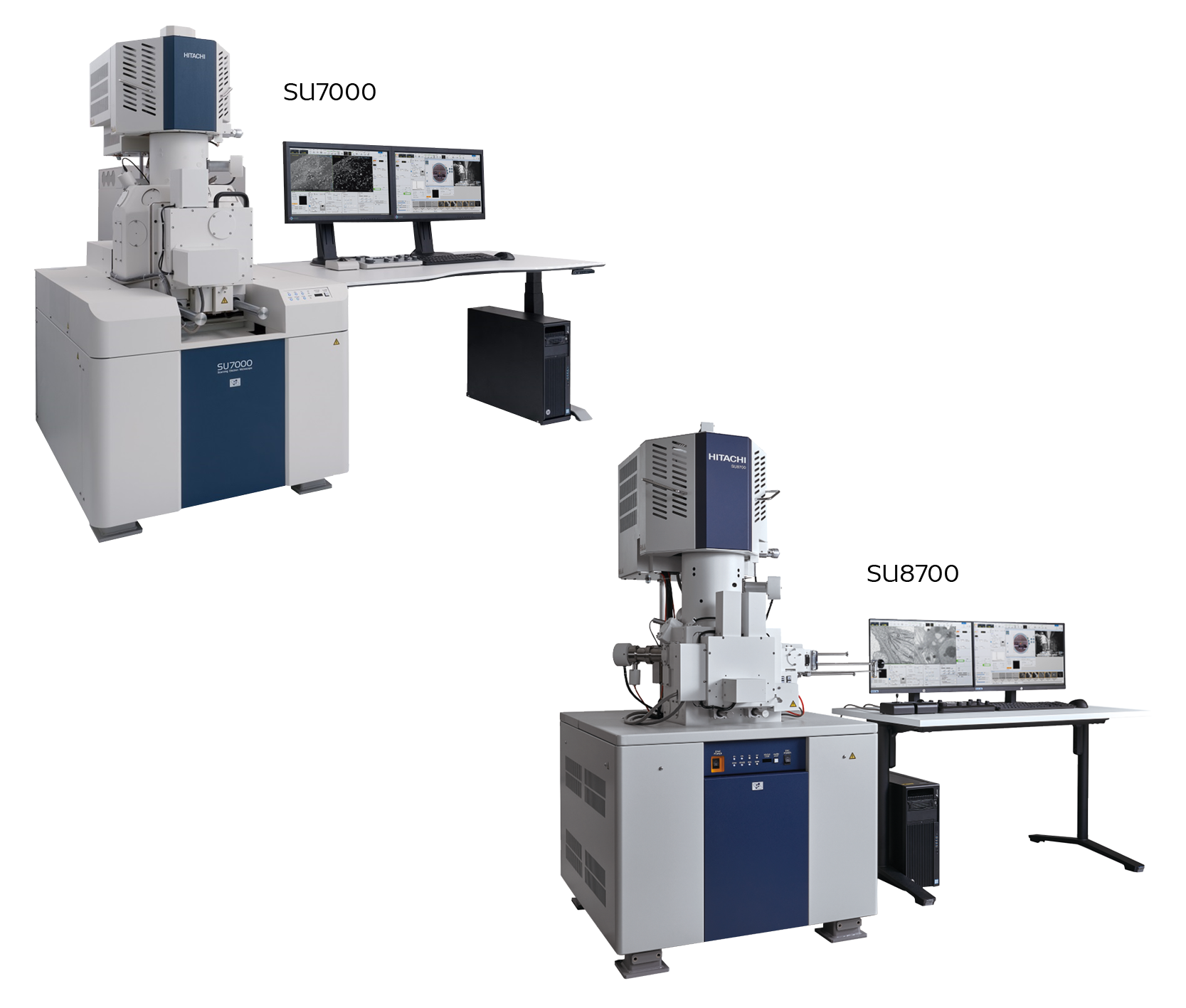
Skip to the main content.
Hitachi_SU7000_SU8700_header image

SU7000 and SU8700 Field Emission SEMs

Schottky FE-SEMs for both ultra high-resolution imaging and analytical work
Common features:
  • Ideal for users who want both ultra high-resolution imaging and analytical capabilities.
  • Display up to six image channels in parallel using a dual monitor user interface.
  • Analytical working distance of just 6 mm.
  • Variable-pressure capabilities without needing extra apertures enable imaging and analytical work on non-conductive specimens.
  • Advanced automation for streamlined workflows.
  • Schottky field emission source. Full range beam boosting and an electrostatic-magnetic hybrid objective lens for sub-nanometer resolution without magnetic sample interaction
SU8700:
  • Optimized for fast (high throughput) inspection of specimens up to 150 mm in diameter.
  • Load samples through a standard six-inch specimen exchange chamber, with an optional inert-gas transfer mechanism.
SU7000:
  • Larger chamber. Accommodates specimens up to 200 mm in diameter and 80 mm in height.
  • 18 chamber access ports, including two cabling ports in the stage door, for optional accessories.

Overview

The SU7000 and SU8700 FE-SEMs from Hitachi offer an exceptional balance between ultra high-resolution imaging and analytical capabilities. Designed for both, these two instruments share the same high-performance electron optics and analytical features, but come with different chamber and stage sizes.

The SU7000 and SU8700 are built to handle a wide range of applications. Their high brightness Schottky field emission electron source, single-scan multi-signal imaging ability, and optional workflow automation (using the EM Flow Creator software) ensure consistent results and superior productivity.

Key Features and benefits

Specifications Table

  SU7000 SU8700
Image Resolution 0.8 nm @ 15 kV, 0.9 nm @ 1 kV 0.6 nm @ 15 kV, 0.8 nm @ 1 kV, 0.9 nm @ 0.3 kV
Magnification 20× to 2,000,000×
Electron Optics - Emitter Schottky Emitter
Electron Optics - Accelerating Voltage 0.1–30 kV (≥0.01 kV optional)
Probe Current Max. 200 nA
Detectors - Standard UD (Upper Detector), MD (Middle Detector), LD (Lower Detector)
Detectors - Optional PD-BSED (Semiconductor Type), UVD (Ultra Variable Pressure Detector), Bright Field STEM Detector, HAADF STEM Detector
Variable Pressure (VP) Mode - Range 5–300 Pa
Variable Pressure (VP) Mode - Available Detectors PD-BSED, UVD, UD, MD, LD
Specimen Stage - Control 5-axis Motor Drive
Specimen Stage - Movable Range (X, Y, Z, T, R) X: 0–135 mm, Y: 0–100 mm, Z: 1.5–40 mm, T: -5–70°, R: 360° X: 0–110 mm, Y: 0–110 mm, Z: 1.5–40 mm, T: -5–70°, R: 360°
Specimen Chamber - Size Max. Ø200 mm, Max. 80 mm Height Max. Ø150 mm
Monitor (Optional) 23-inch LCD (1920×1080), supports dual monitors
Image Display Modes Large Screen (1280×960), Single Image (800×600), Dual Image (800×600, 1280×960), Quad Image (640×480), Hex Image (640×480)
Image Data Saving - Pixel Size 640×480, 1280×960, 2560×1920, 5120×3840, 10240×7680
Optional Accessories EDX, WDX, EBSD, CL, Cryogenic Transfer System, Compatible with various sub-stages EDX, WDX, EBSD, CL, inert-gas specimen transfer

Applications gallery

Applications_SU7000_SU8700_1
Nanoparticles Containing Core-Shell Structure

Fine surface structure is visible by SE signal (UD).

Applications_SU7000_SU8700_2
Nanoparticles Containing Core-Shell Structure

Core (Ag) and Shell (SiO2) are easily distinguishable by BSE signal (MD)

Applications_SU7000_SU8700_3
Voltage Contrast Images of 7 nm process SRAM


Applications_SU7000_SU8700_4
Planview Image of 3D NAND Flash Memory


Applications_SU7000_SU8700_5
Ultrastructure of Arabidopsis thaliana


Applications_SU7000_SU8700_6
Ultrastructure of Arabidopsis thaliana

For Energy Filtered BSE detection, ultrastructure such as thylakoid membrane are clearly visible

“I would definitely recommend Hitachi to other
interested parties and would also choose Hitachi
again in a subsequent decision.”

BIOTRONIK representative, user of SU7000 SEM and ArBlade 5000

Biotonik Logo

Speak to our experts

Our specialists are here to guide you in selecting the right SEM for your needs. Reach out to learn which microscope is best for you: The SU7000 or the SU8700. You’ll also learn how either can enhance your lab’s imaging capabilities, streamline workflows, and support your scientific or industrial applications.


Contact us today to learn more about the SU7000 and SU8700 FE-SEMs.