4 min read
Your Questions Answered: Electron Microscope Workflows with EM Flow Creator
Aug 7, 2026, 11:16:56 AM
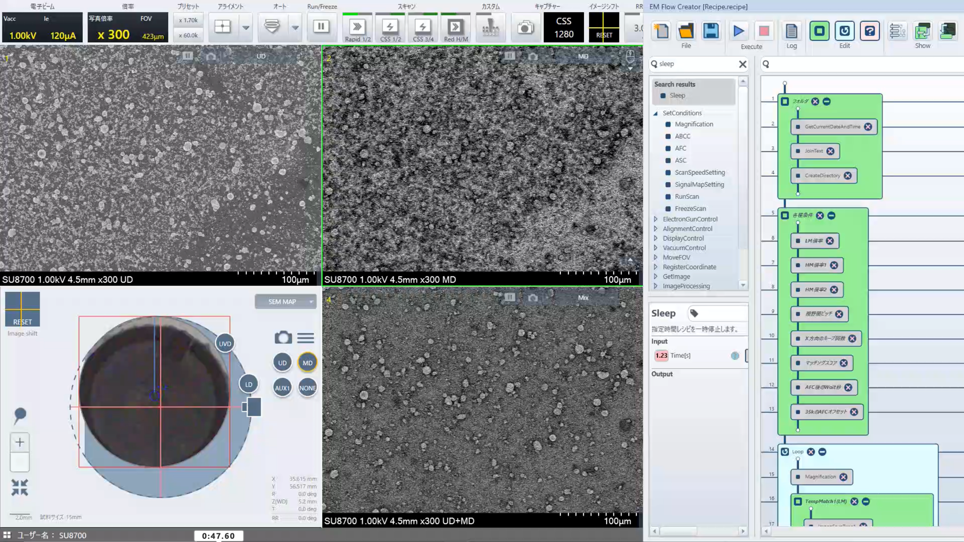
Repetitive acquisition work on a TEM or SEM ties skilled people to a microscope for hours, doing something the instrument could do on its own. EM Flow Creator (EMFC) is Hitachi's visual scripting software that solves this problem: you build a recipe by arranging blocks on screen rather than writing code, and the microscope runs it.
We recently ran a live webinar on automated electron microscopy, covering how recipes are built, how automation handles real samples that don't behave uniformly, and where it fits into standards-driven work like asbestos analysis and technical cleanliness.
The questions that came in were specific and mostly practical. Here are the answers.

Which electron microscopes are supported by EM Flow Creator?
EM Flow Creator is available across the current Hitachi range:
- Tabletop series: Including the TM4000PlusIII and FlexSEMII
- Tungsten SEMs: Including the SU3800 and SU3900
- Field emission SEMs: Including the SU3900SE, SU3800SE, SU8700, SU7000, and SU8600
- TEMs: Including the HT7800 family; HF5000
The available command set differs between them, based on the software platform and the optical systems. TEM recipes work with a different set of blocks than SEM recipes.
There's also a version specifically for the tabletop series, covering routine automatic image acquisition without the functions that aren't relevant on a tabletop instrument.
Is 100 images the maximum a recipe can acquire?
No, 100 was just the example shown in the webinar. The ceiling comes from your microscope's control software, not from EMFC. Some instrument software versions cap out at 9,999 images.
What types of measurements can the software perform?
EMFC is an acquisition and automation tool, not a measurement package, though basic measurements can be performed.
What it controls is the sequence:
- Accelerating voltage
- Magnification
- Detector selection
- Stage movement
- Auto brightness and contrast
- Auto focus and stigma correction
- Loops
- Conditional logic
- Image processing
- Python script blocks if you need them
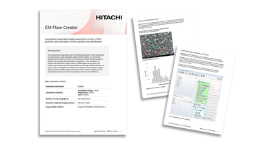
In the particle sizing example shown during the webinar, EMFC acquired the image set and passed it to image analysis software that handled binarisation and particle size distribution automatically.
Can we access raw detector signals during image acquisition? For example, current measured on the BSE detector?
EMFC reads out the detector exactly as it would if you pressed capture, and saves in whatever formats the capture command supports on your instrument. If your microscope only writes TIFF, JPEG and BMP, that's what EMFC writes. If it supports a raw format, EMFC can save raw. The same applies to 8-bit versus 16-bit.
How does the software handle focus, stigmation, brightness and contrast when moving across a large sample? If the sample isn’t perfectly horizontal, that could be a problem.
You control how often correction happens. Auto brightness, auto focus, and auto stigmation can each be called during the recipe at whatever interval suits the sample.
That flexibility matters because the right answer depends on your sample and your preparation. A well-mounted flat specimen might rarely need correction. An inclined or topographically varied surface might need it often.
Does the software support automatic detection of spots on a filter, and navigation to each one, even when their positions are irregular and there's no consistent inter-spot distance?
Yes. This is what target and template matching is for: the software detects the feature itself, so irregular spacing and unpredictable positions don't matter.
The image processing block covers more than template matching alone. Binarisation and histogram modification are in there too, which means you can tune fairly precisely what does and doesn't count as a target. Setting that up takes more effort than a simple particle or fibre target, but it's front-loaded effort. Once the recipe works, it works every run.
Is template recording and image cropping now part of EM Flow Creator, or does it still have to be done manually outside?
Cropping still has to be done manually, outside EMFC. That step exists so you can confirm the correct target has been selected before the recipe starts working from it.
Is there smart template matching that could recognise, say, a triangular particle within a size tolerance, at any orientation on screen?
Not yet. As AI is integrated into later versions, that's a plausible route, but it isn't available today.
For including EDS in the workflow, with Oxford Instruments AZtec, for example, does EM Flow Creator control the AZtec software?
EMFC starts the software and runs the measurement, but it can't control Oxford-specific settings. Those need to be saved in a profile beforehand; otherwise the default or last-used settings apply.
EDS is generally better integrated than other third-party software, because SEM and EDS already communicate bidirectionally about stage position, magnification and so on.
Which EDS vendors can be used through EM Flow Creator? Are there requirements for the EDS software version?
At the current development stage, Oxford Instruments is the only vendor to have supplied the full command list needed to address EDS properly. Work is under way with other vendors, including Bruker, to reach the same level of integration.
Version and module requirements depend on the combination of microscope, EDS detector and software version, so that's a question for your Hitachi contact rather than one with a universal answer.
Key Takeaways from the Webinar
- Bridges simple tools and full coding: EM Flow Creator is a visual automation layer for Hitachi EMs, combining drag‑and‑drop workflows with optional Python scripting for advanced users.
- Automates repetitive imaging at scale: Handles multi-magnification, multi-detector, and tiled “snake” imaging to cover large areas or many samples quickly. Ideal for particle analysis, asbestos, and cleanliness workflows.
- Improves consistency and reproducibility: Standardizes imaging conditions (focus, brightness, stigmation, magnification) so large datasets are captured under tightly controlled, repeatable parameters.
- Enables unattended and overnight runs: Orchestrates batch imaging across multiple filters or positions, freeing operators from low‑value manual scanning and allowing labs to use off‑hours microscope time.
- Integrates with analysis and EDS tools: Integrates with compatible third‑party software such as Media Cybernetics Image Pro and Oxford AZtec EDS, turning raw image capture into end‑to‑end automated workflows.
Related Resources