What our application experts think about the TM4000 benchtop microscopes
Patrick Marks is a Senior Technical Applications Specialist for Electron Microscopes and Specimen Preparation Equipment at Hitachi High-Tech Europe with over 23 years of experience working with our microscopes. We recently spoke to him about Hitachi's TM4000 Benchtop Scanning Electron Microscopes (SEMs) and the many benefits they provide for customers.
Could you give us a quick overview of the TM4000 Benchtop SEMs?
The TM4000Plus Benchtop Electron Microscope is a compact, portable electron microscope which offers all the advantages of a standard SEM such as high-resolution imaging and the ability to provide material composition and topographical information. It also has the benefit of being able to conduct advanced analytical work such as full EDS analysis/mapping and automated particle analysis that normally would only be possible with a full-sized SEM instrument.
What benefits can customers expect when using one of the Benchtop TM4000 Microscope range?
With the TM4000Plus, samples need minimal preparation before imaging. Another excellent benefit is that uncoated samples can be imaged thanks to the microscope’s Variable Pressure function, allowing users to take their samples directly from their experimental process or production environment and place them in the microscope.
The TM4000Plus also has a unique Variable Pressure Secondary Electron (SE) detector that allows the SE signal to be obtained under low vacuum conditions, something a standard SE detector cannot do. Additionally, the TM4000Plus has very low power requirements, lower running costs, lower cost of ownership, and lower environmental impact.
What sectors and industries can benefit from using the TM4000 Microscope series?
It is very difficult to pinpoint a specific target market for the TM4000Plus. This microscope is the widest ranging, most versatile, flexible instrument available that covers an incredible range of applications for academia and all manner of industries alike, from Biomaterials and Polymers through to Electronics, Materials and Metallurgy.
The ease of use and flexibility of the TM4000Plus has made it a leader in academia for students and researchers alike to get results quickly and easily, while similarly, the ability to obtain and feedback results to the relevant manufacturing process or R&D team means lower downtime and higher cost savings for many companies in multiple industries.
Quite simply, anybody who requires higher resolution than is possible with an optical microscope or those who need more in-depth information from their samples such as elemental analysis and so forth can benefit from the powerful capabilities of the TM4000Plus.
How does the TM4000 series compare to other market competitors?
The microscopes have many features such as Secondary Electron imaging in low vacuum conditions, wide area image acquisition that allows automated multiple image capture over a wide area and subsequent stitching into a large, single image. Additionally, users can change vacuum settings in real time whilst imaging the sample.
The low vacuum SE detector allows the TM4000 series to perform both STEM (Scanning Transmission Electron Microscopy) and CL (Cathodoluminescence) imaging, which is a very powerful capability.
Additionally, the TM4000 series has a powerful SEM MAP function. An optical image of the sample is captured upon sample loading, which is then overlaid with an EM thumbnail of the sample when a micrograph is captured. This allows users to obtain a sample navigation record that can be exported for reports.
The TM4000 series boasts great ease of use, with an extremely easy to master user interface and simple routine maintenance, which minimizes downtime. Furthermore, for analytical needs, Hitachi work with Oxford Instruments and Bruker Nano Analytics, both world leaders in Surface Metrology and Analytics.
Finally, as an Electron Microscopy expert, what is your favourite feature of the TM4000Plus and why?
There are many features I like on the TM4000Plus. Firstly, the ability to see and collect both Backscatter Electron (BSE) and Secondary Electron (SE) signals at the same time, combined with the ability to mix the two signals together (simultaneous SEM and BSE imaging), allows the best of both worlds – all while in low vacuum mode.
Other outstanding features are the versatility of the Low Vacuum SE detector which allows both STEM and CL imaging, the Wide Area Image Acquisition and Image stitching function, and the TM4000Plus’s overall flexibility and universal imaging ability which allows the instrument, and user, to image virtually anything.
Revolutionise your SEM sample preparation
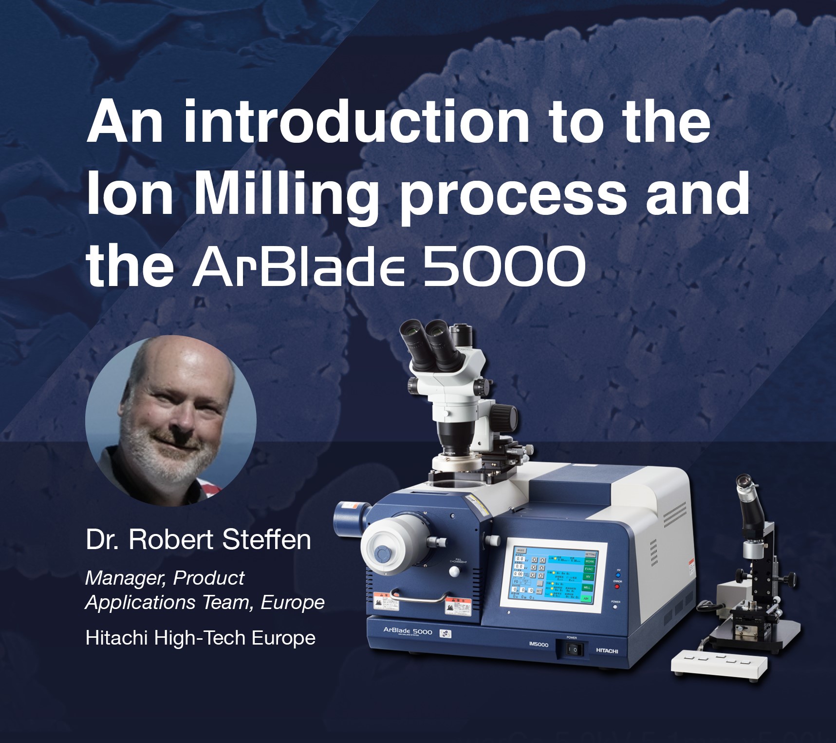
In this on-demand video, Dr. Robert Steffen introduces Broad Ion Beam Milling: the innovative technique that produces defect-free, millimeter-order smooth specimen surfaces for further analysis, even on highly complex and sensitive specimens.
Topics from this blog: TM4000 Products Benchtop SEM
Back